Your visual field
At our office we do an automated visual field test on every patient "old enough" to do it. Generally this means teenagers and older. Recently I've been telling patients why we do it. I'm surprised at how many people just didn't know. I also am surprised that most eye exams don't include such a valuable test. A visual field test, especially an automated visual field, is essential for maintaining not only healthy eyes but a healthy brain.
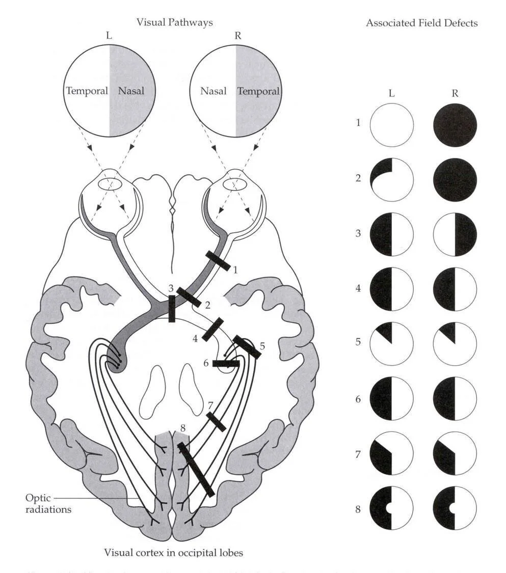
visual field damage and associated neurological region
The visual pathway extends throughout the brain. Many eye doctors (yes both ophthalmologists and optometrists) will not perform an automated visual field at every exam. At our office we set an exceptionally high standard on your health. We include this visual field as part of our comprehensive exam. In recent times alone we have seen visual field loss that looks like this:
Patient A:
Healthy 49 year old male. Here for my annual eye health check up. No complaints.
the patients visual field results. The black areas in both eyes indicate a problem.
This patient had an undiagnosed pituitary tumor. An MRI was ordered and he ended up with surgery. The tumor was completely removed and his visual fields are now normal.
Patient B:
70 year old male. He came in for an emergency visit. His chief complaint "something seems wrong with my vision but I don't know what for the last 2 hours"
can you see the small black spot in the middle in both eyes...that is a problem
The fact that the defect is the same in both eyes is indicative of something going on in the brain. This patient was sent to the ER and was treated for a stroke.
Patient C
35 year old male with a chief complaint. "I think I have a convergence issue. The same one you diagnosed my family member with."
this is what we call an early bilateral superior temporal defect
The defect is very subtle. After seeing his visual field I asked if any hormonal changes had taken place in the last few months. He told me recent lab work had indicated his thyroid was under-active. The patient was sent for an MRI and an early pituitary tumor was found.
Patient D
15 year old male. "I have trouble copying from the board at school"
this one is a deep superior left side defect
The patient was sent for an MRI. Atrophy was noted in his parietal lobe. It was suspected to be from trauma.
Patient E
60 year old female. "I have headaches and no one can tell me why."
this one is a small inferior left side defect
The patient was sent for an MRI and a small stroke was found.
Some of the photos may be hard to see. You as the reader may not even understand what the photos mean. Luckily we do. So as you can see visual field loss can be "silent." Most people don't have notable visual field complaints. As you can also see automated visual fields should be the standard at every eye exam. It is a tool often reserved only for glaucoma but can be a valuable asset to any office. Please come in and be a part of the most comprehensive eye care team. Send your loved ones in too. His or her "eyesight" might be fine but vision is so much more than that.


